
Ottawa Ankle Rules
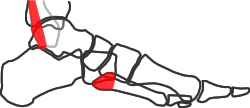
Tenderness over:
- posterior half of lateral malleolus
- base of 5th metatarsal
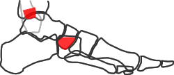
Tenderness over:
- posterior half of medial malleolus
- navicular tuberosity
or unable to bear weight in emergency department:
Xray is indicated
The standard plain radiographic views of the injured ankle are the mortise view and lateral. The mortise view is a modified AP with the ankle internally rotated so that the malleoli are in the same horizontal plane and the joint space is seen evenly on both sides of the ankle. This requires 10-20deg internal rotation. Adding a true AP view does not add useful information (Vangsness 1997, Brage 1997), and should not be part of the standard series.
The mortise view is taken in internal rotation, so that the malleolar tips are in the same horizontal plane – the amount of rotation varies between patients but is usually 10-20deg. A true lateral is very valuable in assessing congruence and posterior malleolar fractures.
We obtain ankle Xrays weightbearing when possible, but in the acute trauma situation the patient usually cannot comply fully.
The Ottawa ankle rules (Stiell et al 1994) have been shown to be accurate in predicting the need for radiography in the acute trauma situation. They can be used accurately by medical and nursing staff in a variety of settings, and can reduce unnecessary radiography. However, like many decision aids, their main function may be to ensure the practitioner carries out a proper clinical assessment.
CT can be useful in assessing more complex fractures. Black (2013) found that CT changed management in 40% of syndesmotic injuries, 31% of fracture-dislocations and 29% of trimalleolar fractures. Scans influenced fixation of medial and posterior malleolar fracture elements.
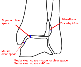
Key measurements on the mortise view of the ankle
Measurements on ankle radiographs
Displacement of an ankle fracture refers to the alignment of the talus in the mortise. This is normally identified according to the medial clear space. The medial clear space is normally no more than the superior clear space. However, at least 4mm and probably 5mm of medial clear space normally indicates an intact DDL and a stable ankle (Koval 2007). Minor degrees of fibular rotation are irrelevant if the mortise is congruent:
- in stable fractures with an intact deltoid ligament, the distal fibular fragment is congruent with the talus and the mortise is congruent; the apparent external rotation is due to internal rotation of the proximal fragment by muscle action (Magid et al 1992, Harper 1995). However, in unstable injuries with a deltoid tear or medial fracture, the distal fibular fracture is truly externally rotated and the medial clear space is increased (Tang et al 2003).
- the talus does not “follow the fibula” (Yablon 1977) unless the DDL is torn or the medial malleolus detached from the tibia (Michelson 1996)
Murphy (2012) measured medial and superior clear space in 73 patients without ankle injuries. 17% of male Xrays and 1% of female Xrays had a medial clear space >4mm, while 2% of males and no females had a medial clear space >5mm. 13% of radiographs had a medial clear space greater than superior clear space. Measurements were symmetrical, so the authors suggest the use of contralateral comparison radiographs to evaluate apparent medial widening.
Radiographic assessment of the syndesmosis is discussed in the page on syndesmotic injuries.
Stress radiographs
Several papers (McConnell 2004, Egol 2004, Schock 2006) have suggested that external rotation stress Xrays can differentiate stable from unstable undisplaced fractures. Most of these used manual applica- tion of stress, but Schock used a “gravity stress view” with the patient in the lateral decubitus position, and reported this was less painful than manual stress but equally sensitive. These papers cast doubt on the accuracy of clinical signs in predicting instability – however, their main outcome measure was talar displacement under external rotation, with 4mm as the upper limit of normal. Schuberth (2004) showed this was a poor predictor of arthroscopically-diagnosed DDL tears.
If instability is defined as “abnormal movement under normal load”, normal loading of the ankle is primarily axial rather than rotational. Weber (2010) reported the use of unprotected standing radiographs as stress tests, with 9% of 56 undisplaced fractures displacing under load. Akhtar (2009) reported 153 patients with clinical evidence of potential instability in which protected weightbearing views showed displacement in 2%.
Koval (2007) investigated the significance of positive stress radiographs with MR imaging of the deep deltoid ligament. They found that 91% of patient with positive stress radiographs had only partial deltoid tears and were successfully treated in a walking boot.
Hoshino (2012) reported a study of weightbearing radiography in 36 patients who had undisplaced malleolar fractures, medial tenderness, bruising or swelling and positive external rotation stress radiographs. Only three showed increased medial joint space on weightbearing radiography; the other 33 were successfully treated non-surgically. Tornetta (2012) treated 54 stress-positive fractures in cast after discussion with patients (60 chose surgery). All 54 healed without displacement as measured by weightbearing medial clear space. These studies support Weber’s (2010) conclusion that stress radiographs greatly over-estimate instability and that most undisplaced ankle fractures can be treated non-surgically.
To summarise these findings:
- If it’s displaced, it’s unstable
- If it’s undisplaced, it’s probably stable
- No medial tenderness, bruising or swelling >99%
- Medial tenderness, bruising or swelling 90-98%
- Stress Xrays probably over-estimate true instability